The upper gastrointestinal tract comprises the oesophagus, stomach and duodenum (duodenum) - the organs that are examined endoscopically during an oesophago-gastro-duodenoscopy (OGD). This complex anatomical region presents a variety of diagnostic and therapeutic challenges: from precise navigation through the tortuous structures to the detection of subtle pathological changes and the safe performance of minimally invasive procedures.
Many diseases in this area have an unfavorable prognosis if they are discovered late. For example, the 5-year survival rate for esophageal cancer is only around 24-25 % overall, because only around a third of tumors are diagnosed at an early stage - although survival rates of up to 75 % are possible with early diagnosis.
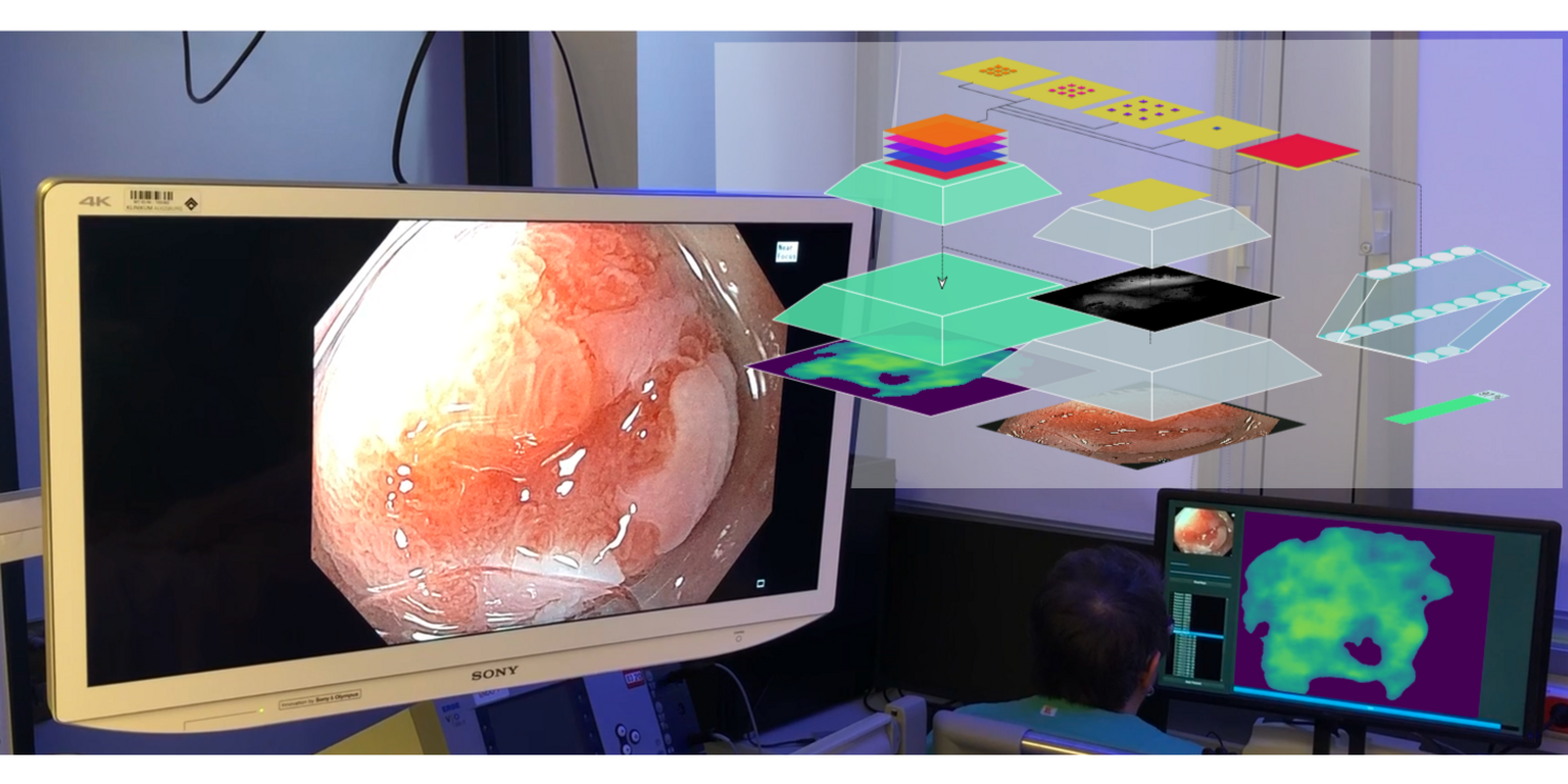
This is precisely where our projects come in: We are developing AI-supported systems that support doctors in early detection, precise surgical planning and intraoperative decision-making - for faster, safer and more effective treatments.